
What Are Dog Ultrasounds?
An ultrasound exam (ultrasonography) is a non-invasive imaging technology used to examine a dog’s internal organs and soft tissues.
An ultrasound machine uses sound waves to create an image of a part of your dog’s body. Ultrasound is used to examine the structure and movement of organs and can also be used to evaluate blood flow in an area.
An ultrasound exam is a common form of imaging performed in dogs, second only to x-rays.
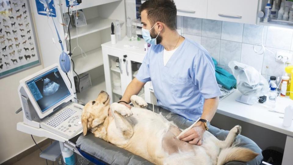
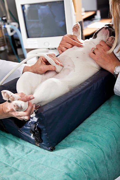
To perform an ultrasound exam, the veterinary professional places a probe on your pet’s body surface and evaluates the image that is transmitted to the screen.
There’s a lot of training involved in performing an ultrasound and the images can be saved for the veterinarian to review, or they can be sent to a specialist for evaluation.
To improve the image quality, the area of the dog’s body being scanned is usually shaved, and then ultrasound gel is applied. The gel provides better contact between the probe and the skin, allowing the sound waves to pass into the body without passing through air.
An ultrasound machine uses sound waves to create an image of a part of your dog’s body. Ultrasound is used to examine the structure and movement of organs and can also be used to evaluate blood flow in an area. Some dogs rest comfortably for an ultrasound scan, but it’s not uncommon for them to need sedation if the scan is of a large area, such as the entire abdomen.
Types of Dog Ultrasounds
Abdominal ultrasound: An abdominal ultrasound can be used to search the abdomen for abnormalities, or it can be performed on an emergency basis to detect bleeding inside the abdomen, or the presence of a cancerous mass.
Echocardiogram: An echocardiogram is an ultrasound exam of the heart. This diagnostic test evaluates the heart chambers, blood flow through the heart, and the sac that encloses the heart (pericardial sac).
Single-organ ultrasound: A single-organ ultrasound is limited to one structure. For instance, a dog with bloody urine may have a bladder scan, or a pregnant female dog may have her uterus scanned.
Thoracic ultrasound: Thoracic ultrasound is used to look at the heart and the space around the lungs. Fluid can accumulate in this space due to trauma, cancer, infectious causes or heart failure. Fluid can also build up around the heart (inside the pericardial sac) and cause pacing of the heart (arrthmyas) leading to fainting episodes or even death. This is a very quick, non-invasive and useful test, that can be performed even if the dog is having trouble breathing because it can be done while the dog is sitting or standing.
Soft-tissue ultrasound: Soft-tissue ultrasound is used to evaluate muscles, tendons, and ligaments, and is mostly useful in sports medicine.
Why Would a Dog Need an Ultrasound?
A dog may need an ultrasound for many reasons, such as:
- Traumatic injury: An ultrasound may be used to look for free fluid in the abdomen, which could suggest issues such as internal bleeding or a ruptured bladder after a dog was hit by a car.
- Cancer: Ultrasound can be used to identify masses within the abdomen, enlarged lymph nodes, and other changes that may suggest the pet has cancer. Free fluid can also be found in the abdomen in case of a ruptured tumor in the spleen or the liver.
- Chronic vomiting and/or diarrhea: The gastrointestinal tract may be scanned to look for causes of vomiting or diarrhea.
- Urinary tract issues: The bladder and kidneys can be scanned for stones, crystals and masses, or be used to collect a urine sample.
- Pregnancy: The uterus can be scanned to diagnose pregnancy, determine the age of the fetuses, and monitor fetal development and viability.
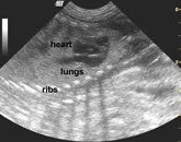
We can see the ribs (the white structure in the middle causing shadowing because ultrasound waves do not go through bone), the developing heart and lungs of the fetus.
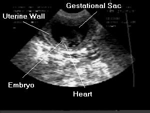
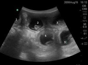
This picture shows 5 embryos inside each placental sac (the black area is the fluid around the stem embryo) and the placenta supplies nutrition and oxygen for the developing puppies.
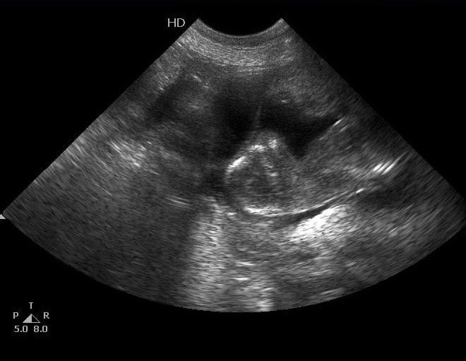
This picture shows a more developed puppy inside the uterus. We can see its head and his chest (he’s on his side). The black area is the fluid inside the placenta, which protects the fetus from movement on the outside of the body.
Ultrasound pregnancy confirmation is possible as early as day 20-22, but can be missed this early. Ultrasound at day 30 post- mating is a great diagnostic tool to confirm pregnancy. Ultrasound has the drawback of not being accurate to count puppies, but can determine heartbeats in the puppies.
- Heart disease: An echocardiogram is the standard test for diagnosing and monitoring heart disease.
- Abnormal lab work or chronic illness: Ultrasound may be used to identify changes to organs that could explain abnormal lab results or chronic illness. For example, the liver can be enlarged in cases of progressive heart disease, infection or cancer. The adrenal gland could be enlarged in some dogs with Cushing’s disease or the pancreas will be brighter in cases of pancreatitis (due to the severe inflammation).
Risks of Dog Ultrasounds
An ultrasound carries minimal risk. Sound waves are not dangerous, and no radiation is involved.
There is a risk of bleeding if an ultrasound is used for guiding biopsies. For instance, if an abnormality is seen in the liver, a needle can be inserted into the abdomen guided by the ultrasound to collect a sample from the abnormal tissue.
Other risks include skin irritation from shaving or gel application, and complications from sedation (if needed).
Benefits of Dog Ultrasounds
The benefits of dog ultrasounds include:
- Real-time imaging
- Non-invasive technique
- Often performed without sedation
- No radiation exposure
- Detailed images for assessing soft tissues
- Ability to monitor blood flow in an organ or a large blood vessel with the ultrasound unit’s Doppler capabilities.
- Effectiveness of Dog Ultrasounds
Dog ultrasounds are highly effective for looking at the structure and movement of soft tissues and most organs.
However, they’re not useful for evaluating air-filled organs such as healthy lungs or the stomach. In addition, they’re not useful for imaging bones or tissues encased in bone, such as the brain or spinal cord, because sound waves cannot pass through bone.
Cost of Dog Ultrasounds
A dog ultrasound usually costs between $300 and $600. Quick one-organ scans or pregnancy diagnosis scans typically cost less. The cost may increase if your dog is large or requires sedation.
Preparation for Dog Ultrasounds
Ultrasounds for dogs don’t usually require at-home preparation, other than withholding food and water for 8 to 12 h if possible (not recommended in young puppies). If there’s food in the stomach, it can cause artifacts and make it difficult to find delicate structures, such as the pancreas.
Written by Rhiannon Roehler, DVM
Adapted by Luana Torres, DVM